Looking for a Tutor Near You?

Post Learning Requirement » x
Ask a Question
x

Choose Country Code

x

Direction

x

Ask a Question

x

Hire a Tutor

Notes On Importance Of Protozoa

Published in: Biology
1,050 Views

Point form with illustration and diagram make students easy to understand the concept.

Jenn Y / Melaka

10 years of teaching experience

Qualification: Master

Teaches: English, Chinese Mandarin, Accounts, Mandarin, Mathematics, Additional Math, Modern Maths, Flute, Keyboard, Cello, Music Theory, Biology, Accounting, Additional Maths, Math

Contact this Tutor
  1. 2_17 _ Ll 8 Importance of protozoa 1. 2. 3. 4. 5. 6. 7. 8. Parasitic protozoa are usually small, have short generation times, high rates of reproduction and a tendency to induce immunity to reinfection in those hosts that survive. Feeding - parasitic protozoa feed can feed using a variety of mechanism such as phagocytosis, pinocytosis and endocytosis. Respiration - Protozoa have examples of both aerobic (Plasmodium) aerotolerant and anaerobic respiration (Amoeba). Reproduction a. Asexual: by simple division such as Amoeba and Giardia; Multiple division; Schizogony (repeated nuclear division followed by cytoplasmic division within the original cell or schizont) Coccidia; b. Sexual: associated with reduction division and fusion of gametes with subsequent chromosomal recombination. Mastigophora, Ciliophora, Amoebas: some parasitic forms. Apicomplexa are all parasitic Globally some of the most important disease-causing agents in human and animals Phylum Sarcomastigophora (with flagella, pseudopodia, or both} Apicomplexa (apical complex) 00 Subphylum Mastigophora (flagella) Sacodina (pseudopodia) Microspora Ciliophora (with cilia) Unclassified (((((ITO 00 Representative Genera Leishma.')ia Trypanosoma Trichomonas Cotamoeya Oieotamoeba and Acanthamocba Babesia Toxop;asma Enterocytozoon Bat'antidium Pneumæystis Major Diseases Produced in Human Beings Visceral. cutaneous and rnucocutaneous infection S! eepertg sickness Chagas' disease Diarmea Vaginitis Dysentery. liver abscess Coli Its Central nervous system and corneal ulcets Babesiosie Malaria Diarrhea Diarrhea Diarrhea Toxopfasmosis Diarrhea Dysentery Pneumonia Entamoebahistolytica a. b. c. d. e. f. g. h. Cysts are passed in feces(l). Infection by ingestion of mature cysts(2) fecallycontaminated food/water/ hands. Excystation (3) in the small intestine and trophozoites(4) are released-migrate to large intestine. The trophozoitesmultiply by binary fission-produce cysts(5)-passed in the feces. Because of the protection conferred by their walls, the cysts can survive days weeks (transmission). It has been established that the invasive and noninvasive forms represent two separate species, respectively E. histolyticaand E. dispar, however not all persons infected with E. histolyticawill have invasive disease; morphologically indistinguishable. •ymptoms of "amoebiasis"-intermittent and mild (various gastrointestinal upsets, including colitis anddiarrhea). In more severe cases the gastrointestinal tracthemorrhages, resulting in dysentery. In some cases thetrophozoiteswill enter the circulatory system and infect other organs, most often the liver (hepaticamoebiasis), or penetrate the gastrointestinal tract -Y acute peritonitis; such cases are often fatal. Infection often diagnosed by demonstrating cysts ortrophozoitesin a stool sample.
  2. i. j. Worldwide distribution, with higher incidence of amoebiasisin developing countries. In industrialized countries, risk groups include travelers and recent immigrants, and institutionalized populations. Infective Stage Diagnostic Stage mature cysts ingested Passed in feces Noninvasive Colonization Intestinal Disease É.ttraintestinal Disease Exits host Mudipbcation Trophozoites o Cysts 9. Trypanosomes a. b. c. d. e. f. Trypanosomacruzi, causes Chagasdisease, a zoonoticdisease that can be transmitted to humans by blood-sucking triatominebugs. An infected triatomineinsect vector ('kissing' bug) takes a blood meal and releases trypomastigotesin its feces near the site of the bite wound.Trypomastigotesenter the host through the wound or through intact mucosalmembranes, such as the conjunctiva(l). Inside the host, the trypomastigotesinvade cells, where they differentiate into intracellular amastigotes(2). The amastigotesmultiply by binary fission(3)and differentiate into trypomastigotes, and then are released into the circulation as bloodstreamtrypomastigotes (4). Trypomastigotesinfect cells from a variety of tissues and transform into intracellular amastigotesin new infection sites. Clinical manifestations can result from this infective cycle. The bloodstream trypomastigotesdo not replicate. Replication resumes only when the parasites enter another cell or are ingested by another vector. The 'kissing' bug becomes infected by feedingon human or animal blood that contains circulating parasites (5). The ingested trypomastigotestransform into epimastigotesin the vector's midgut(6). The parasites multiply and differentiate in the midgutand differentiate into infective metacyclictrypomastigotesin the hindgut (7,8). T.cruzican also be transmitted through blood transfusions, organ transplantation, transplacentally, and in laboratory accidents. Geographic Distribution:the Americasfrom the southern US to southern Argentina. Mostly in poor, rural areas of C & S America. ChronicChagasdisease is a major health problem in many Latin American countries. With increased population movements, the possibility of transmission by blood transfusion has become more substantial.
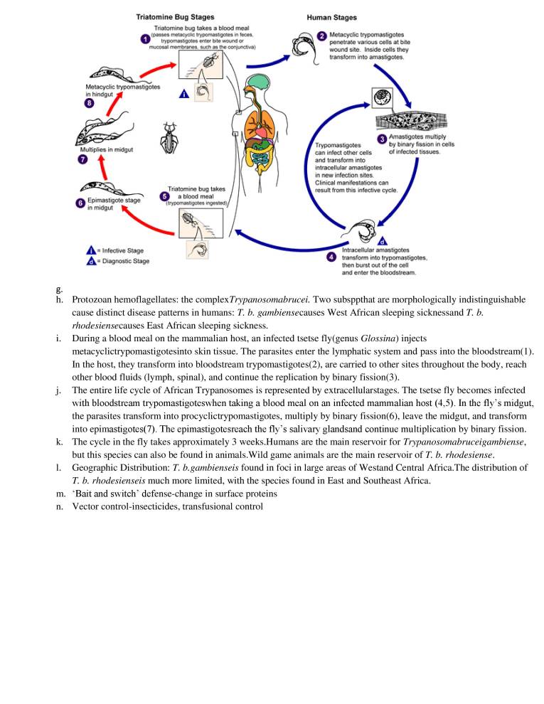
  3. Triatomine Bug Stages Triatomine bug takes a blood meal rr•etaeyebe in O trycornasbootes Ode or *ugh aorvuneiva) Metacyclic trypomastigotes in hindgut Multiplies in midgut O Épimastigote Stage in midgut Infegtvve Stage Diagnostic Stage Triatomine bug takes a blood meal Human Stages O Metacyclic trypomasligotes penetrate various cells at bite wound Site. Inside cells they transform into amasligobes, O Arnastigoies multiply by binary fission in cells Trypgmastigates can infect other Cells of infected tissues, and transform into intracellular amastigotes •n new infection sites. Clinical manifestations can result from this infective cycoe. Intracenular arnastigotes O transform into trypomastigötes„ then burst out of the cea and enter the bloodstream, g. h. i. j. k. l. m. n. Protozoan hemoflagellates: the complex Trypanosomabrucei. Two subsppthat are morphologically indistinguishable cause distinct disease patterns in humans: T. b. gambiensecauses West African sleeping sicknessand T. b. rhodesiensecauses East African sleeping sickness. During a blood meal on the mammalian host, an infected tsetse fly(genus Glossina) injects metacyclictrypomastigotesinto skin tissue. The parasites enter the lymphatic system and pass into the bloodstream(l). In the host, they transform into bloodstream trypomastigotes(2), are carried to other sites throughout the body, reach other blood fluids (lymph, spinal), and continue the replication by binary fission(3). The entire life cycle of African Trypanosomes is represented by extracellularstages. The tsetse fly becomes infected with bloodstream trypomastigoteswhen taking a blood meal on an infected mammalian host (4,5). In the fly's midgut, the parasites transform into procyclictrypomastigotes, multiply by binary fission(6), leave the midgut, and transform into epimastigotes(7). The epimastigotesreach the fly's salivary glandsand continue multiplication by binary fission. The cycle in the fly takes approximately 3 weeks.Humans are the main reservoir for Trypanosomabruceigambiense, but this species can also be found in animals. Wild game animals are the main reservoir of T. b. rhodesiense. Geographic Distribution: T. b.gambienseis found in foci in large areas of Westand Central Africa.The distribution of T. b. rhodesienseis much more limited, with the species found in East and Southeast Africa. Bait and switch' defense-change in surface proteins Vector control-insecticides, transfusional control
  4. Tsetse fly Stages epimasligotes multiply in salivary gland, They transform into metacyclic trypomastigotes. Procyclic trypomastigotes leave the midgut and transform into epimastigotes. Bboodstream 'trypomastigotes transform into procyclic trypomastigotes in tsetse fly's midgut. Procyclic tryposmatjgoteS multiply by binary fission. O. 10. Plasmodium spp. Tsetse 'fly takes a blood meal (injects tryponusbgetesj Tsetse fly takes a blood meal are ingested) Human Stages Injected metacyclic trypomastigotes transform into bloodstream trypomastigotes. which are carried to other Sites, Trypomastigotes multiply by binary fission in various body fluids, e.g.. blood, lymph, and spinal fluid, o Trypomastigotes in blood Infective Stage Diagnostic Stage a. b. c. d. e. f. g. h. i. j. k. l. m. Suborder Haemosporina, Family Plasmodiidae i. Parasites of vertebrate tissue and blood cells ii. Transmitted by blood sucking insects Malaria-'bad air' Bacillus; 1884-Plasmodium i. I .5b endemic areas; over 500 million infected; 1-2 million deaths per year Approx. 156 named sppof Plasmodium which infect various species of vertebrates. Four are known to infect humans: P. falciparum, P. vivax, P. ovaleand P. malariae Malaria generally occurs in areas where environmental conditionsallow parasite multiplication in the vector- Anopheles- 1902 Nobel prize usually restricted to tropicaland subtropicalareas and altitudes below 1,500 m. Distribution might be affected by climatic changes, egglobal warming, and population movements. Both P. falciparumand P. malariaeare encountered in all shaded areas of the map (P. falciparum, P. vivaxby far the most prevalent). stable or endemic malaria: sustained incidence over several years, includes seasonal transmission, various levels of endemicity, immunity and disease tolerance correlate with endemicity, epidemics unlikely unstable or epidemic malaria: marked increase in incidence, population is non-immune, morbidity and mortality can be high Population lacking Duffy blood group-resistant to P. vivaxmalaria-90% west african Clinical manifestations- i. cold stage: feeling of intense cold, vigorous shivering, lasts 15-60 minutes ii. hot stage: intense heat, dry burning skin, throbbing headache, lasts 2-6 hours iii. sweating stage: profuse sweating, declining temp, exhausted, weak sleep,lasts 2-4 hours The malaria parasite life cycle involves two hosts. During a blood meal, a malaria-infected female Anopheles mosquito inoculates sporozoites into the human host (1). Sporozoites infect liver cells(2) and mature into schizonts(3), which rupture and release merozoites(4). (NB: P. vivax and P. ovalea dormant stage [hypnozoites] can persist in the liver and cause relapses by invading the bloodstream weeks, or even years later.) After this initial replication in the liver (exo-erythrocyticschizogony), the parasites undergo asexual multiplication in the erythrocytes (erythrocyticschizogony). Merozoites infect rbc(5). The ring stage trophozoites mature into
  5. n. o. p. q. r. s. t. u. schizonts, which rupture releasing merozoites(6). Some parasites differentiate into sexual erythrocytic stages (gametocytes)(7). Blood stage parasites are responsible for the clinical manifestations of the disease. The M (microgametocytes) F (macrogametocytes), are ingested by an Anopheles mosquito during a blood meal(8). The parasites' multiplication in the mosquito = sporogonic cycle. While in the mosquito's stomach, the microgametes penetrate the macro gametes generating zygotes(9). The zygotes in turn become motile and elongated (ookinetes)(10) which invade the midgut wall of the mosquito where they develop into oocysts(l l). The oocysts grow, rupture, and release sporozoites(12), which make their way to the mosquito's salivary glands. Inoculation of the sporozoites into a new human host perpetuates the malaria life cycle. Infective stages: i. ookinete(motile): mosquito gut epithelial cells ii. sporozoite(motile): mosquito salivary glands, hepatocytes iii. merozoite(non-motile): erythrocytes Prevention and control: i. ii. iii. iv. lhuman-mosquito contact reduce vector—DDT; resistance reduce parasite reservoir vaccine Treatment —quinine, chloroquine-resistant strains Re-emergence: 1950s Kenya Mosquito Stenos cell Human Liver Stages Cycle Release of "POTOZ0ites Sportnonic Cyclo 000kinete Macrogemgtogyte Miaogamete enterirg rnacrogamele O Éxfjageilated m•crogametøcyte Infective Stage Diagnostic Stage Mosquibo takes a blood meal (injects o Mosquito takes {ingests P m.Wariw Ruptured sehizgnt o Schizont Human Blood Stages ln•Mnature trophoaode €1 Erytuocytic Cyee ring Stage J Mature Ruptured schizot'it Schizgnt Gametocytes 11. Symbiotic protozoa a. b. Flagellatesare important parts of zooplankton,life that drifts near the surface of large bodies of water, where they form the baseof many aquatic food chainsand may be producing most of the atmosphere's oxygen. i. Amoebaeconstituted a major component of the heterotrophic nanoplanktonicassemblage ii. Tintinnids, a type of Ciliate, are common in the marine plankton Sedimentation: Foraminiferansand radiolariansof the phylum ciliata. Foramsare encased in compartmented calcareouscoverings, while the radiolarian coverings are ornately sculptured and covered with delicate spines. Pseudopodia, many with skeletal elements, radiate from the cell and catch diatoms and small protozoa. After death, their coverings accumulate on the sea floor contributing to the bottomsediments.
  6. c. d. e. f. g. h. Water pollution indicator: fecal pollutionEg. GiardiaandCryptosporiumspp. (resist Cl) -risk to human health MutualismSymbiont: i. ii. iii. iv. v. vi. Trichonymphaagilisinhabits the gutof common eastern termite, Reticulitermesflaviceps Anaerobic, lack mitochondria and chloroplasts Trichonymphaenters the host (termite) by way of other termites ingesting adult termite fecal matter The termites would starve without its endosymbionts. Termites will chew and ingest wood, but lack the enzymes.For this they depend on their microbial guests. In return, the termite provides a favorable, safe and stablemicroenvironmentand a steady supply of raw materials. Nutrient cycling-mineralising nutrients ( N—>NH4+) Grazing of bacteria-control, stimulate growth Food source; compete/feed on pathogens flagella nuc leus wood particle