Looking for a Tutor Near You?

Post Learning Requirement » x
Ask a Question
x

Choose Country Code

x

Direction

x

Ask a Question

x

Hire a Tutor

Notes On Antigen

Published in: Biology
1,169 Views

With the Notes, learn about how antigen works in our immune system. It is cool!

Jenn Y / Melaka

10 years of teaching experience

Qualification: Master

Teaches: English, Chinese Mandarin, Accounts, Mandarin, Mathematics, Additional Math, Modern Maths, Flute, Keyboard, Cello, Music Theory, Biology, Accounting, Additional Maths, Math

Contact this Tutor
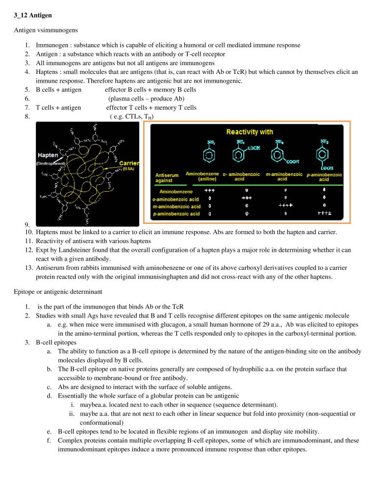
  1. 3_12 Antigen Antigen vsimmunogens 1. 2. 3. 4. 5. 6. 7. 8. 9. 10. 11. 12. 13. Immunogen : substance which is capable of eliciting a humoral or cell mediated immune response Antigen : a substance which reacts with an antibody or T-cell receptor All immunogens are antigens but not all antigens are immunogens Haptens : small molecules that are antigens (that is, can react with Ab or TcR) but which cannot by themselves elicit an immune response. Therefore haptens are antigenic but are not immunogenic. B cells + antigen T cells + antigen Hapten 'h-lv-— effector B cells + memory B cells (plasma cells — produce Ab) effector T cells + memory T cells ( e.g. CTLs, TH) Carrie (B SAI Aminobenzene Antiserutn {aniline} against Aminobenzene o-aminobenzoic acid m-amimobenzoic acid p•aminobenzoic acid Reactivity with o- aminobenzoic acid COOK m•aminobenzoic p-arninobenzoic acid acid Haptens must be linked to a carrier to elicit an immune response. Abs are formed to both the hapten and carrier. Reactivity of antisera with various haptens Expt by Landsteiner found that the overall configuration of a hapten plays a major role in determining whether it can react with a given antibody. Antiserum from rabbits immunised with aminobenzene or one of its above carboxyl derivatives coupled to a carrier protein reacted only with the original immunisinghapten and did not cross-react with any of the other haptens. Epitope or antigenic determinant 1. 2. 3. is the part of the immunogen that binds Ab or the TcR Studies with small Ags have revealed that B and T cells recognise different epitopes on the same antigenic molecule a. e.g. when mice were immunised with glucagon, a small human hormone of 29 a.a., Ab was elicited to epitopes in the amino-terminal portion, whereas the T cells responded only to epitopes in the carboxyl-terminal portion. B-cell epitopes a. b. c. d. e. f. The ability to function as a B-cell epitope is determined by the nature of the antigen-binding site on the antibody molecules displayed by B cells. The B-cell epitope on native proteins generally are composed of hydrophilic a.a. on the protein surface that accessible to membrane-bound or free antibody. Abs are designed to interact with the surface of soluble antigens. Essentially the whole surface of a globular protein can be antigenic i. maybea.a. located next to each other in sequence (sequence determinant). ii. maybe a.a. that are not next to each other in linear sequence but fold into proximity (non-sequential or conformational) B-cell epitopes tend to be located in flexible regions of an immunogen and display site mobility. Complex proteins contain multiple overlapping B-cell epitopes, some of which are immunodominant, and these immunodominant epitopes induce a more pronounced immune response than other epitopes.
  2. inject antiserum anti- anti—e anti-z anti-' 4. 5. 6. 7. antibodies of different specificity heterogeneity affinity isotype h. The antiserum obtained following the injection of a complex antigen such as a protein contains antibodies that recognise many different epitopes. i. These antibodies will also differ in their affinity and isotype. T-cell epitopes a. T cells do not recognise soluble native Ag but instead recognise Ag that has been processed and is presented in association with MHC molecules. b.) Ag processing is required to generate peptides that interact specifically with MHC molecules. c.) Epitopes recognised by T cells are often internal. Antigep presenting cell Agretope MHC Epitope TCR T cell b. Antigenic determinants on Abs a. b. c. Isotypic i. Constant region determinants that distinguish each H & L chain class and subclass ii. isotypes define constant region determinants that distinguish each heavy chain class and subclass and each light chain type and subtype within a species. Each isotype is encoded by a separate constant region gene. Allotypic i. Structurally different alleles of the same gene ii. multiple alleles exist for a particular gene resulting in proteins with different allotypes; structural differences between alleles can give rise to antigenic determinants e.g. human kappa light chains have two alleles one of which has a val at a.a. 191, the other leu. Idiotypic i. Structures unique to a variable region; may be associated with Ag binding site ii. each individual antigenic determinant is an idiotype; the sum of the individual idiotopes is the idiotype of the antibody. Some but not all idiotopes are associated with the antigen binding site. For Abs there is allelic exclusion: the products of only one allele are present within one antibody genes offspring ANTIBODIES NEVER"
  3. 8. 9. What determines if something is effective in eliciting an immune response? a. b. c. d. e. Foreigness i. Must be recognized as "non-self' by the immune system ii. In general the greater the degree of foreignness, the stronger the response Size i. Best immunogens usually at least 100,000 daltons ii. Most (but not all) substances smaller than 5000-10000 Da are poor immunogens Composition and heterogeneity i. Chemical complexity contributes to immunogenicity ii. Homopolymers are less immunogenic than copolymers Are processed and presented i. ii. iii. Both the humoral and the cell-mediated immune response are aided by interaction with T cells. Antigen presenting cells present processed Ag in the context of MHC molecules to activate T cells Molecules that cannot be degraded (eg. D-amino acids) are poor immunogens Treatment to increase uptake by antigen presenting cells improves immunogenicity o Cross-linking o Aggregation o Attachment to insoluble matrices For TCR: ability to be processed & displayed in the MHC Proteins: excellent immunogens 10. Polysaccharides: good — LPS 11. DNA: poor — denature-hapten 12. Lipids: poor — hapten Adjuvants 1. 2. 3. 4. 5. 6. Adjuvants are substances when mixed with an Ag and injected with it, enhance the immunogenicity of the Ag. Adjuvants are often used to boost the immune response when an Ag has low immunogenicity or when only small amounts of an Ag are available. Adjuvant appear to exert one or more of the following effects: a. Antigen persistence is prolonged b. Co-stimulatory signals are increased c. Local inflammation is increased d. The nonspecific proliferation of lymphocytes is stimulated Many contain molecules that signal through the Toll receptors Freund's Adjuvant a. b. c. d. Incomplete: Ag in aqueous solution, mineral oil, & emulsifying agent i. Ag is slowly released Complete: also contains heat-killed Mycobacteria i. A muramyl dipeptide of the mycobacterial cell wall activates M Both result in granuloma formation i. A granuloma is a dense, macrophage-rich mass of cells. Within the granuloma the macrophage are activated. ii. Activated macrophages are more phagocytic and express molecules (class Il MHC and B7) that aid the immune response (enhance TH response) Note: Freund's complete adjuvant more potent than the incomplete form. Alum (Aluminum potassium sulphate) a. b. c. d. e. Cause precipitation Results in Ag persistence Increased size improves phagocytosis Local chronic inflammatory response (granuloma — macrophage rich mass of cells) Alum prolongs the persistence of antigen. When an Ag is mixed with alum, the salt precipitates the Ag.
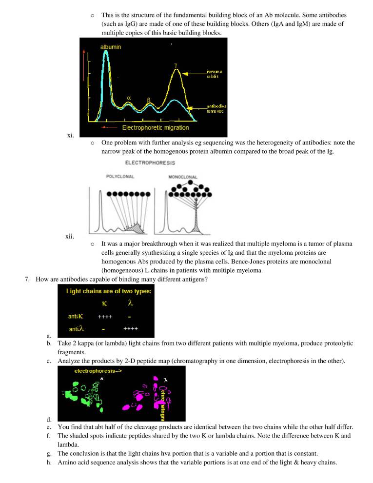
  4. f. g. h. Injection of this alum precipitate results in a slower release of Ag from the injection site, so that the effective time of exposure to the Ag increases from a few days without adjuvant to several weeks with the adjuvant. The alum precipitate also increases the size of the Ag, thus increasing the likelihood of phagocytosis. Note: Alum and Freund's Adjuvant also stimulate a local, chronic inflammatory response that attracts both phagocytes and lymphocytes. Structure and properties of antibodies l. 2. 3. 4. 5. 6. If bacteria are injected into blood, specific substances are found that exhibit different biologic properties a. Agglutinin — specifically clump only the bacteria used for immunization b. Opsonin facilitate engulfment c. Antitoxin — neutralize toxin associated with the immunizing bacteria d. Cytolysis lyse bacteria e. Precipitins from flocculate precipitates when added to supernatant from the bacteria f. (all of these are properties of antibodies) Antibody (Ab) a. A molecule produced by an animal in response to foreign substance (Ag) which has the property of binding the eliciting agent Immunoglobulin (Ig) a. Antibodies are found both as cell-associated receptors (BCR) and as circulating effector molecules Antigen (Ag) a. Any molecule capable of being bound by the combining site of an Ab or the T-cell receptor Immunogen a. A substance capable of eliciting an immune response; all immunogens are antigens but not all antigens are immunogens What is the structure of an Ab? a. Experiment by A. Tiselius and E.A.Kabat (1939) i. ii. iii. iv. v. vi. vii. Immunized rabbits with the protein ovalbumin Divided the immunized rabbits' serum into two aliquots: o Electrophoresis revealed four peaks corresponding to albumin, alpha, beta and gamma globulins. o Reacted with ovalbumin (Ag) and the precipitate formed was removed; the remaining serum which did not react with the Ag was electrophoresed albumin mrrun e blit re rrnued Electrophoretic migration o Immune serum from rabbits before and after the antibodies were removed by precipitation with Ag (ovalbumin). Conclusion: the antibodies migrated as a gamma-globulin. When purified antibodies (produced by dissolving immunoprecipitates) were analyzed in an ultracentrifuge they ran with a 7S sedimentation co-efficient indicating a m.w. of approx. 150,000 0 7S = 150,000 molecular weight Molecular analysis of precipitates showed the valence = 2 Two molecules of Ag were precipitated by each Ab.
  5. o 0 0 Gamma globulin 7S = 150,000 molecular weight Valence = 2 Papain digestion 2Fab + IFc Fab binds Ag but no precipitate FC forms crystals MW of Fab = 45,000 and Fc = 50,000 Ratio: 2 Fab and 1 Fc Fab Eluant Digestion of antibodies with the enzyme papain yielded two fragments, the Fab (fragment antigen binding), & the FC (fragment crystallizable) (Fab')2 pepsin 1 (Fab')2 of 100,000 viii. ix. binds 2 molecules of Ag can precipitate break SS-->2Fab Eluant o Gamma globulin; 7S = 150,000 molecular weight; Valence = 2; Papain 2Fab + IFc 0 Pepsin of 100,000 o FC fragment not recovered from pepsin because it had been digested into multiple fragments Heavy Chain 0 0 Light Chain Elution volume Gamma globulin; 7S = 150,000 molecular weight; Valence = 2; Papain 2Fab + IFc Pepsin of 100,000 Ab+ cleave disulfides 2H(50000) + 2L(25000) Antiserum : Anti-L reacts only with Fab; Anti-H reacts with Fab and Fc; Anti-Fab reacts with H and L If break disulfide bonds and separate on the basis of size you get two products: H and L polypeptides / L chain H chain Reduction Papain Digestion Feb* Feb Pepsi Smell Frsgments H
  6. This is the structure of the fundamental building block of an Ab molecule. Some antibodies (such as IgG) are made of one of these building blocks. Others (IgA and IgM) are made of multiple copies of this basic building blocks. albumin re rmued Electrophoretic migration One problem with further analysis eg sequencing was the heterogeneity of antibodies: note the narrow peak of the homogenous protein albumin compared to the broad peak of the lg. ELECTROPHORESIS xi. xii. POLYCLONAL UONOCLONAL 7. It was a major breakthrough when it was realized that multiple myeloma is a tumor of plasma cells generally synthesizing a single species of Ig and that the myeloma proteins are homogenous Abs produced by the plasma cells. Bence-Jones proteins are monoclonal (homogeneous) L chains in patients with multiple myeloma. How are antibodies capable of binding many different antigens? Light chains are of two types: antLK antLX. a. b. c. d. e. f. g. h. Take 2 kappa (or lambda) light chains from two different patients with multiple myeloma, produce proteolytic fragments. Analyze the products by 2-D peptide map (chromatography in one dimension, electrophoresis in the other). electrophoresis—> You find that abt half of the cleavage products are identical between the two chains while the other half differ. The shaded spots indicate peptides shared by the two K or lambda chains. Note the difference between K and lambda. The conclusion is that the light chains hva portion that is a variable and a portion that is constant. Amino acid sequence analysis shows that the variable portions is at one end of the light & heavy chains.
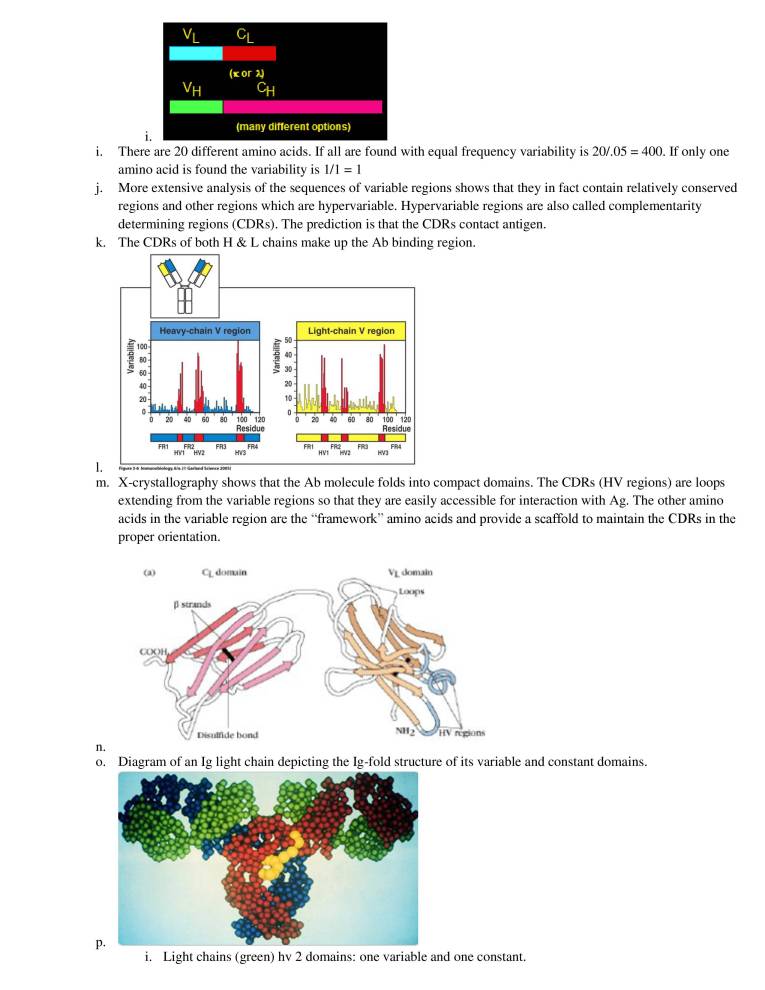
  7. i. j. k. 1. m. n. o. (ror (many different options) i. There are 20 different amino acids. If all are found with equal frequency variability is 20/.05 = 400. If only one amino acid is found the variability is 1/1 More extensive analysis of the sequences of variable regions shows that they in fact contain relatively conserved regions and other regions which are hypervariable. Hypervariable regions are also called complementarity determining regions (CDRs). The prediction is that the CDRs contact antigen. The CDRs of both H & L chains make up the Ab binding region. Heavy-chain V region 100 80 40 20 0 20 40 60 FRI FR2 HVI HV2 50 40 30 20 10 80 100 120 Residue FR4 HV3 Light-chain V region 0 20 40 60 80 100 120 Residue FRI FR2 FR3 FR4 HVI HV2 HV3 Figure 3-6 Immunobiology, Ole. (O Garland Science 2005) X-crystallography shows that the Ab molecule folds into compact domains. The CDRs (HV regions) are loops extending from the variable regions so that they are easily accessible for interaction with Ag. The other amino acids in the variable region are the "framework" amino acids and provide a scaffold to maintain the CDRs in the proper orientation. CL domain strands Disulfide bond VL Oonuin HV n•pons Diagram of an Ig light chain depicting the Ig-fold structure of its variable and constant domains. i. Light chains (green) hv 2 domains: one variable and one constant.
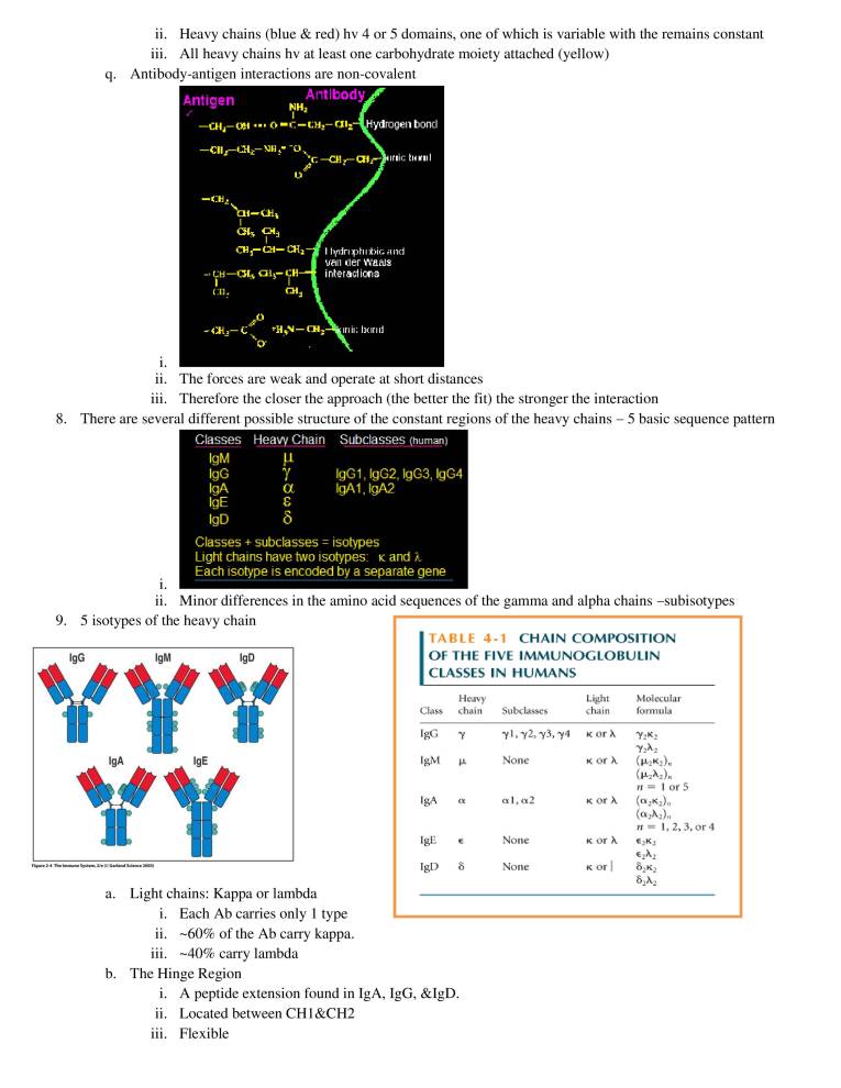
  8. q. ii. Heavy chains (blue & red) hv 4 or 5 domains, one of which is variable with the remains constant iii. All heavy chains hv at least one carbohydrate moiety attached (yellow) Antibody-antigen interactions are non-covalent Antigen Cd—CSI, Antlbody ydrogen boncl c Init; I :phl lid van (ler h'aa]s ims i. ii. The forces are weak and operate at short distances iii. Therefore the closer the approach (the better the fit) the stronger the interaction There are several different possible structure of the constant regions of the heavy chains 5 basic sequence pattern Classes Heaw Chain Subclasses (human) 8. 9. YYY lgM lgG lgE lgD u lgG1, lgG2, lgG3, lgG4 lgA1,lgA2 Classes + subclasses = isotypes Light chains have two isotypes: K and Each isotype is encoded by a separate gene i. ii. Minor differences in the amino acid sequences of the gamma and alpha chains —subisotypes 5 isotypes of the heavy chain CHAIN COMPOSITION TABLE 4-1 OF THE FIVE IMMUNOGLOBULIN CLASSES IN HUMANS lgA Figure 2-4 The Immune System. 2/e (o Garland Science 2005) lgE Class lgA IgE Heavy chain ö Subclasses y 1 , •y2y •y3, Non e al, cg2 None None Light chain R or K or X K or Molecular formula n = I or 5 n = l, 2, 3, or 4 •ö2K2 a. b. Light chains: Kappa or lambda i. Each Ab carries only I type ii. —60% of the Ab carry kappa. • • • . —40% carry lambda 111 The Hinge Region i. ii. iii. A peptide extension found in IgA, IgG, &IgD. Located between CHI &CH2 Flexible
  9. (a) I)NP ligand&—• Hingc region N02 25Å —Anti-DNP s. DNP ligand Ag-Ab Trimer N02 y; Angle between arms is 600 00 Angle between arms is 00 Angle between arms is 900 iv. Hinge is flexible & allows for multitude of Ab-Ag complexes. 10. The levels of Ig before & after birth: fraction of adult level of serum immunoglobulins 100 0 conception —6 Figure 7-20 The Immune System, 2/e (C Garland Science 2005) passively transferred maternal lgG -3 months transient low lgG newly synthesized levels IgM newly synthesize lgG newly synthesized lgA birth3 69 1 2345 adult years b. c. d. e. 5-10% of total serum. Usually the first Ig made in response to antigen. Monomeric IgM, is expressed as membrane-bound antibody on B cells and IgM is secreted by plasma cells as a pentamer.; H chain has 4 constant region domains & no hinge In addition to H & L also contains J(joining) chain which is required for polymerization of the monomers to form pentamers (H2L2)5J or (H2L2)6 Increased valence (10 antigen binding site) leads to increased avidity. 11. Affinity versus Avidity a. b. 12. lgG a. b. c. d. e. f. g. Affinity: strength of one Ab binding site bound to a single antigenic epitope. i. Strength of noncovalent interactions. ii. Low affinity Ab: binds Ag weakly; dissociates more quickly iii. High affinity Ab: binds Ag tightly; remains bound longer. Avidity: strength of multiple interactions between a multivalent Ab& multivalent Ag. i. The affinity of 1 Ab binding site to 1 epitope does not reflect the true strength of an Ab-Ag interaction. ii. High avidity can compensate for a low affinity. Most abundant serum immunoglobulin — 80% of total Ig Subtle amino acid differences between subclasses affect biological activity: i. IgG1,IgG3, and IgG4 readily cross the placenta and play an impt role in protecting the developing fetus ii. IgG3 is the most effective complement activator iii. IgGl and IgG3 bind with high affinity to FC receptors on phagocytic cells and thus mediate oponisation. Most abundant Ab in blood, lymph & extracellular fluids in tissues — produced in plasma cells located in LN, spleen, & BM. Small size: easier access to extracellular spaces of tissue; crosses the placenta. Flexible arm contributes towards IgG having more effector functions than IgM. IgG is transported across the endothelium: Brambell Receptor (FcRB) on endothelial cells. Placenta contains an FcRB receptor to transport IgG to the developing fetus.
  10. FcRB carries lgG across endo- thelium into extracellular spaces extracellular spaces endothelial cell lumen of capillary FcRB lgG 13. lgA a. b. c. d. 14. lgE a. b. i. Figure 7-18 The Immune System, 2/e (o Garland Science 2005) Monomer: plasma cells in the LN, spleen, & BM secrete IgA into the blood. Dimer: plasma cells lining the mucosal associated lymphoid tissue. Dimeric lgA J chain 2-30 Th. Systo-n. 2005) Characteristics of IgA: i. ii. iii. iv. Monomer IgA is found in the blood, lymph, & extravascular spaces. Dimeric IgA is found on mucosal membranes & in secretions: o Milk, saliva, tears, sweat & mucus 0 10-15g of IgA is produced each day; most is secreted. Transcytosis: epithelial cells carry receptor- poly Igreceptor.; binds IgA Fc. Poly Ig Receptor bind the CH3 domain of the FC Binding of IgA to receptor on baso- lateral face of epithelial cell epithelial cell poly-lg receptor dimeric lgA lgA-secreting cell Receptor-mediated endocytosis of IgA tight junction basement membrane Transport of IgA to apical face of epithelial cell lumen lamina propria Receptor is cleaved, IgA is bound to mucus through the secretory piece IgA dimer + secretory component Figure 7-17 The Immune System, 2/e (O Garland Science 2005) Present at extremely low concentrations Responsible for allergy immediate hypersensitivity reactions eg: hay fever, asthma, hives & anaphylactic shock
  11. c. d. e. f. 15. lgD a. Binds to receptors on the membranes of blood basophils & tissue mast cells, will result in the translocation of granules to the plasma membrane & release their contents — gv rise to allergic reactions. Monomer form; Basophils, eosinophils& Mast cells FC Receptors Mast cells: i. Connective tissue: along the blood vessels (dermis of the skin) ii. Of the gastrointestinal tracts & respiratory tract Resting mast cell O O lgE antibody o o 00 ee Resting mast cell has preformed granules containing histamine and other inflammatory mediators Activated mast cell •C oo Multivalent antigen crosslinks lgE antibody bound at the mast-cell surface, causing release of granule contents iii. Figure 7-26 The Immune System, 2/e (O Garland Science 2005) Eosinophil attacking a schistosome larva Together with IgM, is the major membrane bounding Ig expressed by mature B cells. Priciple role seems to be as a membrane lg.; No biological effector function has been identified for IgD. 16. All isotypes can exist as both secreted and membrane bound antibodies a. All isotypes can exist as both secreted & membrane bound antibodies. b. The 2 forms differ in their carboxy terminal sequence; Secreted antibodies hv a hydrophilic terminus. c. Membrane antibodies hv a hydrophobic sequence which inserts into the plasma membrane & a short cytoplasmic tail. Function Neutralization Opsonization Sensitization for killing by NK cells Sensitization of mast cells Activation of complement system Property lgM lgD lgG1 lgG2 lgG3 lgG4 lgA lgE lgM lgD lgG1 lgG2 lgG3 lgG4 lgA lgE Transport across epithelium + dimer Transport across placenta Diffusion into extravascular sites Mean serum level (mg ml-I ) 17. Figure 2-29 The Immune System, 2/e (O Garland Science 2005)