Looking for a Tutor Near You?

Post Learning Requirement » x
Ask a Question
x

Choose Country Code

x

Direction

x

Ask a Question

x

Hire a Tutor

Notes On Nervous System

Published in: Biology
380 Views

How the nervous system works

Ronnie K / Kuala Lumpur

18 years of teaching experience

Qualification: M.Sc.

Teaches: Biology

Contact this Tutor
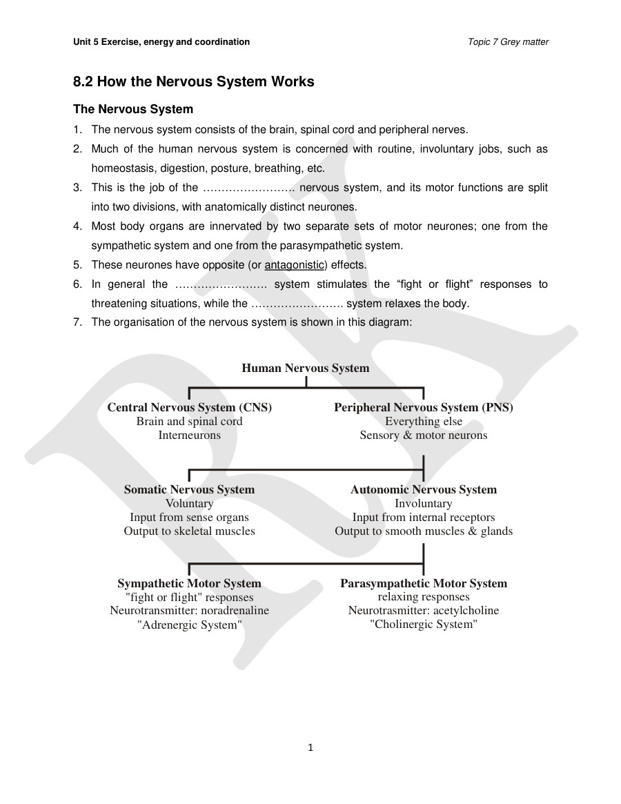
  1. Unit 5 Exercise, energy and coordination 8.2 How the Nervous System Works The Nervous System Topic 7 Grey matter 2. 3. 4. 5. 6. 7. . The nervous system consists Of the brain, spinal cord and peripheral nerves. Much Of the human nervous system is concerned with routine, involuntary jobs, such as homeostasis, digestion, posture, breathing, etc. This is the job of the nervous system, and its motor functions are split into two divisions, with anatomically distinct neurones. Most body organs are innervated by two separate sets of motor neurones; one from the sympathetic system and one from the parasympathetic system. These neurones have opposite (or antagonistic) effects. In general the . system stimulates the "fight or flight" responses to threatening situations, while the — system relaxes the body. The organisation of the nervous system is shown in this diagram: Human Nervous System Central Nervous System (CNS) Brain and spinal cord Interneurons Somatic Nervous System Voluntary Input from sense organs Output to skeletal muscles Sympathetic Motor System "fight or flight" responses Neurotransmitter: noradrenaline "Adrenergic System" Peripheral Nervous System (PNS) Everything else Sensory & motor neurons Autonomic Nervous System Involuntary Input from internal receptors Output to smooth muscles & glands Parasympathetic Motor System relaxing responses Neurotrasmitter: acetylcholine "Cholinergic System" 1
  2. Unit 5 Exercise, energy and coordination Topic 7 Grey matter Sensory, Relay and Effector Neurones . The nervous system composed Of — (adapted to carry nerve impulses) and neuroglia (structural and metabolic support to neurones). Basic structure of a neurone: 2. 3. 4. 5. 6. 7. 8. 9. axon terminal dendron of next neurone A neurone has a direction of nerve impulse axon may be 3' m Eng with extensions leading off it. The cell body contains the nucleus and granular cytoplasm (perikaryon), due to dense clusters of RER (Nissl substances). Numerous . and . . provide a large surface area for connecting with other neurones, and carry nerve impulses towards the cell body from specialised receptors or from adjacent neurones. A single long carries the nerve impulse away from the cell body, either to other neurones or effectors such as muscles. Most neurones have many companion cells called ., which wrap their cell membrane around the axon many times in a spiral to form a thick insulating lipid layer called the — Axons which are covered in myelin are said to be myelinated. Between adjacent Schwann cells, there are gaps where the axons are exposed (uncovered), called . 2
  3. Unit 5 Exercise, energy and coordination 10. Humans have three types of neurone: Topic 7 Grey matter . neurones have long axons and transmit nerve impulses from sensory receptors all over the body to the central nervous system. These are pseudo unipolar neurones with a single dendrite and axon branching from a common stem from the cell body. b) . neurones also have long axons and transmit nerve impulses from the central nervous system to effectors (muscles and glands) all over the body. These are multipolar neurones with dendrites branching from the cell body. c) Bipolar neurones with only a single dendrite arising directly from the cell body opposite the axon. These act as receptors. [Note: Another type Of neurone, interneurones (also called connector neurones or relay neurones) is usually much smaller cells, with many interconnections.] 11. Nerve impulse can be passed from the axon of one neurone to the dendron of another at a synapse. 12. A nerve is a discrete bundle of several thousand neurone axons which may be all sensory, all motor or a combination of both. 13. A nerve fibre is the axon of an individual nerve cell which may carry impulses to (sensory) or from (motor) the brain but not both. 3
  4. Unit 5 Exercise, energy and coordination motor n dend rites body_ o myelin sheath intern many fibres dendrites Cell body nucleus o myelin Sheath Of nor-ve fibros formed by Schmnn cell itself around the Topic 7 Grey matter dendrites Cell body junct»on between two sheath • of Rarwier 4
  5. Unit 5 Exercise, energy and coordination The Nerve Impulse Topic 7 Grey matter 1. Neurones and muscle cells are electrically excitable cells, which means that they can transmit electrical nerve impulses. 2. These impulses are due to events in the cell membrane. The Membrane Potential 2. 3. 4. 5. 6. 7. 8. 9. All animal cell membranes contain a protein pump called the This uses the energy from ATP splitting to simultaneously pump 3 sodium ions the cell and 2 potassium ions If this was to continue unchecked there would be no sodium or potassium ions left to pump, but there are also sodium and potassium ion . in the membrane. These channels are normally closed, but even when closed, they "leak", allowing sodium ions to leak in and potassium ions to leak out, down their respective concentration gradients. 3Na• outside cell membrane inside closed (leak) NaWATPase ATP ADP p. closed . (leak) The combination of the Na+K* pump and the channels cause a stable imbalance of and K* ions across the membrane. This imbalance causes a potential difference across all animal cell membranes, called the The membrane potential is always negative inside the cell. The pump is thought to have evolved as an osmoregulator to keep the internal water potential high and so stop water entering animal cells and bursting them. Plant cells don't need this as they have strong cells walls to prevent bursting. 5
  6. Unit 5 Exercise, energy and coordination The Action Potential Topic 7 Grey matter 2. 3. 4. 5. 6. A. . In nerve and muscle cells the membranes are electrically excitable, which means that they can change their membrane potential, and this is the basis of the nerve impulse. The sodium and potassium channels in these cells are voltage-gated, which means that they can open and close depending on the voltage across the membrane. The normal membrane potential of these nerve cells is —70mV (inside the axon), and since this potential can change in nerve cells It is called the resting potential. When axon is stimulated by mechanical, chemical, thermal or electrical stimulation, a brief reversal Of the membrane potential, lasting about a millisecond, is produced. This brief reversal is called the action potential: time The action potential has 2 phases called depolarisation and repolarisation. Depolarisation. The stimulus cause the membrane potential to change a little. The voltage gated ion channels can detect this change, In and • • . channels open. This causes Na+ to rush in by diffusion (as the Na+ concentration outside is higher), making the inside of the cell more positive. This phase is referred to as a depolarisation since the normal voltage polarity (negative inside) is reversed (becomes positive inside). 6
  7. Unit 5 Exercise, energy and coordination Topic 7 Grey matter B. 7. 8. Repolarisation. When the membrane potential reaches OV, the potassium channels open, causing to rush out (along its concentration gradient), making the inside more negative agaln. In fact, too many leave that the charge on the inside of the membrane becomes more negative than originally, causing an . channels close, restoring the resting potential. Since this restores the original repolarization starts again, polarity, it is called An action potential can be recorded accurately by inserting a very fine glass microelectrode into an axon. Another electrode records the electrical potential from the outside. The results are shown on an oscilloscope. Outside An electrule, made from a glass pipette pulled toa sharp tip, is filled with an electrical conducting solution.. Inside TWO inside and one outside the axon, detect a difference in electric charge in .and connected Wiff a wire to an amplifier. Outside axon Inside axon memb rane Outside axo n 7 The difference is amplified... .md displayed on oscilloscope. rnV Amplifier Time • The constant difference of —63 mV bet-neen outside ara inside is the resting potential.
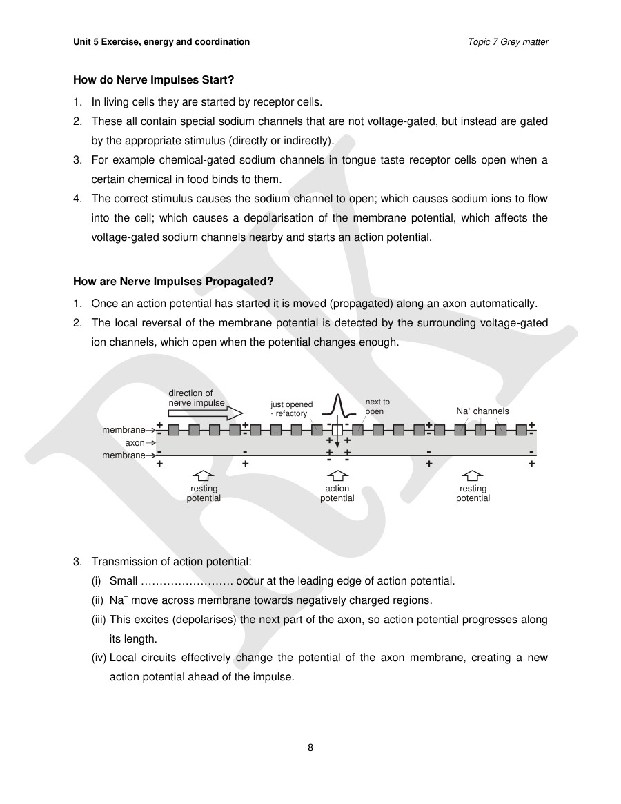
  8. Unit 5 Exercise, energy and coordination How do Nerve Impulses Start? . In living cells they are started by receptor cells. Topic 7 Grey matter 2. 3. 4. These all contain special sodium channels that are not voltage-gated, but instead are gated by the appropriate stimulus (directly or indirectly). For example chemical gated sodium channels in tongue taste receptor cells open when a certain chemical in food binds to them. The correct stimulus causes the sodium channel to open; which causes sodium ions to flow into the cell; which causes a depolarisauon of the membrane potential, which affects the voltage gated sodium channels nearby and starts an action potential. How are Nerve Impulses Propagated? . Once an action potential has started it is moved (propagated) along an axon automatically. The local reversal of the membrane potential is detected by the surrounding voltage gated 2. 3. ion channels, which open when the potential changes enough. direction of nerve Impulse resti ng potential just opened - ref actory next to open membran axon membran action potential Na• channels resting potential Transmission of action potential: (i) Small . . occur at the leading edge of action potential. (ii) Na+ move across membrane towards negatively charged regions. (iii) This excites (depolarises) the next part of the axon, so action potential progresses along its length. (iv) Local circuits effectively change the potential of the axon membrane, creating a new action potential ahead Of the impulse. 8
  9. Unit 5 Exercise, energy and coordination Topic 7 Grey matter 4. The ion channels have two other features that help the nerve impulse work effectively: (i) After an ion channel has opened, it needs a "rest period" before it can open again. (ii) This is called the and lasts about 2 ms. (iii) This means that, although the action potential affects all other ion channels nearby, the upstream ion channels cannot open again since they are in their refractory period, so only the downstream channels open, causing the action potential to move along the axon. (iv) This also limits the . axon. at which successive impulses can pass along (v) The ion channels are either open or closed; there is no half-way position. (vi) This means that the action potential always reaches +40mV as it moves along an axon, and it is never attenuated (reduced) by long axons. (vii)ln other word the action potential is — How Fast are Nerve Impulses? 1. The speed is affected by 3 factors: A. B. C. Axon diameter. • The larger the diameter, the the speed. Myelin sheath. Myelin sheath prevents initiation of action potential / depolarisation. The voltage gated ion channels are found only at the nodes of Ranvier, and between the nodes the myelin sheath acts as a good electrical Action potential / depolarisation only possible at nodes / gaps. The action potential can therefore . node, a process that is called . . . large distances from node to This increases the speed Of conduction (propagation) dramatically, so while nerve impulses in unmyelinated neurones have a maximum speed of around 1 m/s, in myelinated neurones they travel at 100 m/s. Temperature. • The higher the temperature, the faster the speed. • so homoeothermic (warm-blooded) animals poikilothermic (cold-blooded) ones. 9 have faster responses than
  10. Unit 5 Exercise, energy and coordination direction Of myelin node Of Ranvier Sheath -ions channels here only Topic 7 Grey matter 2. 3. 4. 5. 6. 7. 8. 9. In invertebrates, the speed of transmission of a nerve impulse is directly related to diameter of nerve fibre, and there is a limit to how big a nerve fibre can grow. Most vertebrate neurones are associated with Schwann cells and therefore have a myelin sheath. As a result of the nodes of Ranvier, the transmission of a nerve impulse is much faster. So vertebrate nerves that need to carry impulses fast are myelinated, with relatively small diameters. Those that are not myelinated don't need to carry impulses very fast so they can still have small diameters. Squid giant axons are large as they carry impulses relatively quickly during an escape response. This means that they are easy to find and access and easy to insert micropipettes into. Squids are invertebrates, so there are also fewer in experiments of this type. The Synapse . The junction between two neurones is called a synapse. An action potential cannot cross the synaptic instead the nerve impulse is carried by chemicals called . issues with using them between neurones, and 2. 3. 4. 5. These chemicals are made by the cell that is sending the impulse (the pre-synaptic neurone) and stored in synaptic at the end of the axon. The cell that is receiving the nerve impulse (the post synaptic neurone) has . in its membrane (specific binding sites for the neurotransmitters). Roles of enzymes in synapse: (a) They are involved in making the neurotransmitter substances in the presynaptic knobs; (b) involved in the production Of vesicles; (c) involved in the breakdown of neurotransmitters in the synaptic cleft; (d) involved in the production Of ATP to power the various ion pumps and synthesis and breakdown of neurotransmitters. 10
  11. Unit 5 Exercise, energy and coordination mitochondria axon of presynaptic cab o g 90e neurone Sequence of events: Arrival of action potential alters the potential across the . Topic 7 Grey matter dendrite of postsynaptic nurone Na' . membrane. Calcium channels open in the presynaptic membrane, causing calcium ions to flow into the presynaptic knob. These calcium ions cause the 2. 3. 4. 5. 6. 7. . to migrate to the presynaptic membrane and fuse with the membrane, releasing their contents (the transmitter substance / acetylcholine) by exocytosis. The transmitter substance across the synaptic cleft. The transmitter substance binds to the . . in the postsynaptic membrane, causing the Na+ channels to open. Na+ flow in to postsynaptic knob. This causes a . . Of the post synaptic membrane, whereby more Na+ channels open, which may trigger an action potential (when depolarisation reaches . value), also called the excitatory postsynaptic potential. The neurotransmitter is broken down by a specific enzyme in the synaptic cleft; for example the enzyme acetylcholinesterase breaks down the neurotransmitter acetylcholine. This stops the synapse being permanently on. The breakdown products are absorbed by the pre-synaptic neurone by endocytosis and used to re synthesise more neurotransmitter, using energy from the mitochondria. 11
  12. Unit 5 Exercise, energy and coordination 6. 7. 8. 9. Most neurones have many synapses with others. Some are: synapse, ion-specific channels open allowing to enter, to leave. Cause depolarisation of postsynaptic membrane. synapse. nputS from many neurones Topic 7 Grey matter cel body output Neurotransmitter release cause postsynaptic membrane more permeable to K* Produce hyperpolarisation. Balance between the activity of excitatory and inhibitory synapses determine whether action potential is generated. In order for a large enough impulse to be created, summation of impulses can take place: (a) Temporal summation many small impulses summate over time at the . synapse. Each action potential that arrives at the pre-synaptic membrane will cause a number of vesicles to release their transmitter. A number of action potentials are required before there is enough transmitter to initiate an action potential in the post synaptic cell. a) temporal summation presynaptic cell low frequency of action potentials transmitter below threshold high frequency of action potentials transmiter reaches ••• —threshold postsynaptic cell depolarises level no depolarisation of postsynaptic cell 12
  13. Unit 5 Exercise, energy and coordination (b) Spatial summation — many impulses at Topic 7 Grey matter synapses on the same cell allow an impulse to be transmitted, e.g. many rods are needed for one bipolar cell to fire. A number Of pre synaptic neurones may form synapses with one post-synaptic neurone. Action potentials arriving in each pre synaptic neurone will release transmitter, which build up to the threshold level and triggers a postsynaptic impulse. b) spatial summation action potentials only produced action potentials produced in one presynaptic cell no depolarisation of postsynaptic cell TM" inal branch E, Of presynaptic in both presynaptic cells postsynaptic transmitter below threshold potential Threshold of axon of pstsyna*ic Resting (a) Subthreshold, no (b) Tempral surnmation postsynaptic cell depolarises (c) Spatial summation transmitter reaches threshold (d) Spatial summation o' EPSP and IPSP 9. Spatial and temporal summation make an organism more to small stimuli which might not on their own trigger a response. 10. A response coming into several sensory receptors at once, for example, can be added together to give awareness, e.g. the rods of the eye. 11. Similarly, if a small stimulus which would not trigger a post synaptic action potential on its own is repeated several times in quick succession, an organism becomes aware Of it, thus increasing sensitivity and responsiveness. 13
  14. Unit 5 Exercise, energy and coordination How Sensory Receptors Work . The specialized region of the body detecting the . receptor. Topic 7 Grey matter is known as a sensory 2. 3. 4. 5. 6. 7. 8. The simplest and most primitive type Of receptor consists Of a single unspecialized primary . cell composed Of a single sensory neurone whose terminal end is capable of detecting the stimulus and giving rise to a nerve impulse passing to the CNS (e.g. skin mechanoreceptors such as the Pacinian corpuscle). More complex receptors are known as secondary sense cells, and they consist Of modified epithelial cells able to detect stimuli. These form . connections with their sensory neurons which transmit impulses to the CNS (e.g. mammalian taste buds). The most complex receptors are the sensory composed Of a large number of sense cells, sensory neurons and associated accessory structures. In the mammalian eye, there are two types of secondary sense cells, rods and cones, many connecting neurons and many accessory structures such as the lens and iris. On the basis of the position of the receptor and the stimulus, 3 types of receptor are identified: a. b. c. Exteroceptors — these respond to stimuli originating the ear and sound; Interoceptors — these respond to stimuli originating the body, as with the body, such as blood pressure and carbon dioxide receptors in the carotid arteries; Proprioceptors — these respond to stimuli concerned with the relative positions and movements of muscles and the skeleton. An alternative system of classification is based on the type of stimulus detected by the receptor: Type of receptor Type of stimulus energy Electromagnetic Electromagnetic Mechanical Thermal Chemical 14 Nature of stimulus Light Electricity Sound, touch, pressure, gravity Temperature change Humidity, smell, taste
  15. Unit 5 Exercise, energy and coordination 9. Receptors act as biological . responses In axons. Topic 7 Grey matter .., transforming stimulus energy into electrical 10. When a receptor cell receives a stimulus, sodium ions move rapidly across the cell membrane setting up a potential. 11. A small stimulus results in a small generator potential and a large stimulus results in a large generator potential. 12. If the generator potential is large enough to reach the . neurone, an action potential will result in that neurone. 13. If it is not, there will be no action potential. of the receptor 14. In convergence, even if the generator potential from an individual receptor cell is too small to set up an action potential, the generator potentials from several may add together or and trigger an action potential. 15. This makes it possible for the sensory system to respond to stimuli. Muscle Dendrites Cell body hillock 15 Stretching a muscle is the stimulus that activates the opening ot ion channels in stretch receptor dondritos. The resulting depolar- ization spreads to tho cell body, creatinga . .which spreads to the axon hillock, causing action potentials to fire. The action potentials travel down the neuron.
  16. Unit 5 Exercise, energy and coordination Accommodation and Adaptation . Accommodation occurs when . information about in the environment. Topic 7 Grey matter . In a synaptic knob are discharged as a result of too many action potentials in rapid succession. 2. 3. 4. 5. 6. 7. 8. The can no longer respond to the stimulus. Of new vesicles cannot keep up and the neurone A short rest restores the response as new vesicles are made. Accommodation allows organisms to . the CNS does not become overwhelmed with input. . repeated harmless stimuli so that Most receptors initially respond to a strong constant stimulus by producing a high frequency Of impulses in the sensory neurone. The frequency of these impulses gradually decline and this reduction in response, with time, is called adaptation. The advantage of adaptation of sense cells is that it provides the animal with precise At other times the cells remain quiescent, thus preventing with irrelevant and unmanageable information. of the CNS Accommodation: Overstimulation of any presynaptic neurone releases so many synaptic vesicles that further action potentials in the neurone can't release neurotransmitter molecules into the synaptic cleft and no action potential is generated in the postsynaptic neurone. This makes it possible for the animal to concentrate on new, and potentially more important, stimuli. Response returns as new neurotransmitter is synthesised. Adaptation: There is a decrease in the permeability of the receptor membrane to ions due to sustained stimulation. This progressively reduces the amplitude and duration of the generator potential and when this falls below the threshold level the sensory neurons cease to fire (constant stimulation Of receptor cells results in a gradual decline in response). Again this makes it possible for the animal to concentrate on new, and potentially more important, stimuli. Animal will not respond again regardless of amounts of neurotransmitter. 16
  17. Unit 5 Exercise, energy and coordination Accommodation: Accomodation is the response to ignore a stimulus, permanent feature of the environment. if it is a current Topic 7 Grey matter Adaptation: Adaptation is a more response, whereby an increasingly strong stimulus is required for the same response (e.g. the effects of nicotine). Effects of Drugs on Synaptic Transmission . Almost all drugs derive their physiological effect from altering the way synapses work in the brain. 2. By increasing or decreasing the frequency and strength of synapses in different parts of the brain drugs can change mood, perception, memory, judgment and even the ability to co- ordinate actions (e.g. alcohol). 3. Drugs can affect synaptic transmission in various ways, shown in this table: Drug action Mimic a neurotransmitter (i.e. drug has similar shape) Stimulate the release of a neurotransmitter Open receptors on postsynaptic membrane Block receptors on postsynaptic membrane Inhibit the breakdown enzyme Inhibit the Na+K+ pump Block the Na+ or K* channels (i.e. affect permeability pre / post synaptic membranes) Effect Switch on a synapse Switch on a synapse Switch on a synapse Switch off a synapse Switch on a synapse Stop action potentials Stop action potentials Examples Levodopa, nicotine caffeine, cocaine cocaine, caffeine atropine, curare, opioids, atropine neostigmine, DDT tetrodoxin, anaesthetics 4. Drugs that stimulate a nervous system are called agonists, and those that inhibit a system are called antagonists. 17
  18. Unit 5 Exercise, energy and coordination Evidence for the current model of synaptic transmission: Topic 7 Grey matter Electron micrographs: Botulinus toxin: Nicotine. Strychnine etc.: Curare: Show the presence Of vesicles in the synaptic knob Of the presynaptic neurone before an action potential. After repeated action potentials these vesicles are no longer visible, implying that they have released their contents as a result of stimulation. Also shows large numbers of mitochondria that supply the energy for the synthesis of the neurotransmitters etc. the release of acetylcholine and so shows that acetylcholine from the presynaptic membrane is needed for the transmission of an action potential across a synapse. Stimulates the nervous system by binding to the post-synaptic membrane, . the effect of acetylcholine and so suggesting that acetylcholine does the same. Show that acetylcholine causes the setting up of action potentials in postsynaptic neurones, because preventing the breakdown of acetylcholine causes the neurones to respond Shows that blocking of acetylcholine stops transmission of action potentials from nerve cells to muscle cells confirming that acetylcholine needs to bind to acetylcholine initiate post-synaptic potential. 18 to
  19. Unit 5 Exercise, energy and coordination The Reflex Arc Topic 7 Grey matter . The three types Of neurones are arranged in circuits and networks, the simplest Of which is the reflex arc. In a simple reflex arc, such as a withdrawal reflex (e.g. pin prick), a stimulus is detected by a 2. 3. 4. 5. 6. 7. 8. 9. cell, which synapses with a neurone. The sensory neurone carries the impulse from site of the stimulus to the central nervous system (the brain or spinal cord), where it synapses with an . Interneurone allows impulse to pass from sensory neurone to motor neurone. Also allowing Other connections. The interneurone synapses with a . neurone, which carries the nerve impulse out to an . ., such as a muscle, which responds by contracting. Muscle stretch reflexes (e.g. knee jerk) involve only two neurones, sensory and effector. Importance of reflex arc: (b) Increases . response to stimulus chances (as an escape response) (c) Protects body from Reflex arc can also be represented by a simple flow diagram: external stimuli internal stimuli cell or organ Brain or spinal cord (interneurones) 19 muscles or glands movement, secretion, behaviour
  20. Unit 5 Exercise, energy and coordination Detection of Light in Animals The Eye cornea pupil lens aqueous humour suspensory ligaments ciliary muscle ciliary body Topic 7 Grey matter clera horoid etina rteries and veins itreous humour ptic nerve ye muscle The Sclera The Choroid The Retina The strong, tough outer layer (containing collagen fibres) that holds the eye together. Front part is transparent forming the cornea. The spherical shape of the eye is maintained by the pressure of the liquid inside. Curved surface refracts light on to the retina This layer contains the blood vessels that feed every cell of the eye. It also contains the pigmented cells that make the retina appear black (prevents reflection of light within the eye). Modified to form the pigmented iris at the front. This contains the light sensitive photoreceptor cells and their associated neurones supplying the optic nerve. Responds to stimulus Of light by producing action potential leading to production of impulses which are transmitted to the brain via the optic nerve. 20
  21. Unit 5 Exercise, energy and coordination Topic 7 Grey matter The Cornea The Iris The Lens The Ciliary body The Humours This is a specialised part of the sclera at the front of the eye. It is made Of aligned collagen fibres and is transparent and tough. This is made Of pigmented cells, which give eye colour, and radial and circular muscles, which control the amount of light entering the eye. This is a transparent, rubbery tissue made of crystallin proteins, which crystallise to form a biconvex, glass-like lens. The cells are laid down in layers as the eye develops to form the lens shape, but then die, so the lens does not need a blood supply. This supports the lens. It comprises circular muscles and radial elastic fibres called suspensory ligaments. Together theses control the shape of the lens. These are the fluids (transparent, containing gelatinous mucoprotein) inside the eye, secreted by the cells Of the choroid. The vitreous humour behind the lens is more viscous than the aqueous humour in front of the lens. Contribute to the maintenance of the shape of the eye. 21
  22. Unit 5 Exercise, energy and coordination The Retina Topic 7 Grey matter . The retina contains the photoreceptor cells and their associated interneurones and sensory neurones. They are arranged as shown in this diagram: light to c ganglion nerve cells bipolar neurones rod cone pigmented cells cells retina 2. 3. A surprising feature of the retina is that it is back-to-front (inverted). The retina is made up Of 3 layers Of cells: The outermost, photoreceptor layer The intermediate, middle layer The inner layer kik The photoreceptor cells are at the back of the retina, and the light has to pass through several layers of neurones (small and transparent) to reach them. • There are two kinds of photoreceptor cells in human eyes: and . ., both which are partly embedded in pigmented epithelial cells Of the choroids. Contains neurones (special interneurones) which form synapses with rods and cones. Synapse with sensory neurones called . cells. Contains ganglion cells, Of which the axons cover the inner surface of the retina and eventually form the . nerve (containing about a million axons) that leads to the brain. 22
  23. Unit 5 Exercise, energy and coordination Visual Transduction . Visual transduction is the process by which light initiates a nerve impulse. Topic 7 Grey matter 2. 3. 4. Rods and cones act as they convert one form of energy to another form. They convert light energy to electrical energy (nerve impulse). Light stimulus is detected by the photosensitive pigments . (in cones). (in rods) and Rods Rods are evenly distributed throughout the retina but are absent from the fovea. Rods are extremely sensitive to light, but do not discriminate colours. 2. 3. 4. 5. 6. 7. 8. 9. Rods respond to — dim light and night vision. The structure of a rod cell is: light intensities (than cones), so are principally used in inner segment outer segment synapse nucleus mitochondria membrane disks The detection of light is carried out on the membrane disks (membranous vesicles) in the outer segment. These disks contain thousands Of molecules of rhodopsin ('visual purple'), the photoreceptor molecule (pigments absorbing light). The inner segment contains: (a) Numerous mitochondria (provide ATP for rhodopsin resynthesis) (b) Polysomes (synthesis of proteins for the production of visual pigments) (c) Nucleus Rhodopsin responds to all wavelengths of light. Rhodopsin consists Of a membrane-bound protein called covalently bound prosthetic group called . and a 10. Retinal is made from vitamin A, and a dietary deficiency in this vitamin causes night- blindness (poor vision in dim light). 11. Retinal is the light sensitive part, and it can exists in 2 forms: a cis form and a trans form: 23
  24. Unit 5 Exercise, energy and coordination li ht - fast 22. This produces a potential in the rod. sensory neurone in the nerve to the brain. Topic 7 Grey matter Rhodopsin with trans retinal dark - slow (mins) 12. In the dark retinal is in the form, but when it absorbs a photon of light it 13. This changes its shape and therefore it can no longer bind tightly to opsin. Rhodopsin with cis retinal quickly switches to the form. 14. This process is called retinal and opsin. ., whereby rhodopsin splits (or, bleaches) into 15. The reverse reaction (trans to cis retinal) requires an enzyme reaction and is very slow, taking a few minutes. 16. This explains why you are initially blind when you walk from sunlight to a dark room: in the light almost all your retinal was in the trans form, and it takes some time to form enough cis retinal to respond to the light indoors. 17. Rod cell membranes contain a special sodium channel that is controlled by rhodopsin. 18. Rhodopsin with cis retinal opens it and rhodopsin with trans retinal closes it. 19. This means in the the channel is open, allowing sodium ions to flow in. 20. In the light, the rod cell membrane is much less permeable to Na+ as Na+ channels are closed. 21. As the sodium pump continues to pump Na+ out of the cell, the interior becomes more negative than usual 23. If the generator potential is large enough to reach the threshold, or if several rods are stimulated at once, the neurotransmitter (glutamate) is not released into the synapse with the bipolar cell. [Note: The synapse with the bipolar cell is an inhibitory synapse. Glutamate is an inhibitory neurotransmitter.] 24. This sets up an action potential in the bipolar cell which passes across the synapse to cause an action potential in the sensory neurone leading to the brain. 25. The final result of the bleaching of the rhodopsin in a rod cell is a nerve impulse through a 26. In the visual areas of the brain this visual information is converted into an awareness of the Image. 24
  25. Unit 5 Exercise, energy and coordination In The Dar* rod cell bipo ar cell extra toy ganglion cell Cones retinal Na• channels open —xne mbrane depo larise d U neurotransmitter released o inhibitory neuroreceptor open ± membrane hyperpolarised no neurotransmitter released no action potential rod cell bipolar exc tat", y ganglion cell Topic 7 Grey matter In The Light T Na• channels closed —nembrane hyperpolarised U no neurotransmitter released 0 inhibitory neuroreceptor closed ± membrane depolarised (action potential) " neurotransmitter released action potential 2. 3. 4. 5. 6. Although there are far more rods than cones, we use cones most of the time because they have fine discrimination and can resolve colours. To do this we constantly move our eyes so that images are focused on the small area of the retina called the fovea. You can only read one word of a book at a time, but your eyes move so quickly that it appears that you can see much more. The more densely-packed the cone cells, the better the visual acuity. In the fovea of human eyes there are 160 000 cones per mm2, while hawks have 1 million cones per mm2, so they really do have far better acuity. The stimulation of cone cells results in a more detailed image (vs. rod cells) as: a) b) c) d) e) One cone linked to one ., thus One cone stimulated, one impulse to brain Several rods linked to one ganglion / bipolar cell / convergence Several rods stimulated to trigger one nerve impulse Information from several rods is combined / pooled (summation) 25
  26. Unit 5 Exercise, energy and coordination Rods and Cones Topic 7 Grey matter 1. Why are there two types of photoreceptor cell? The rods and cones serve two different functions as shown in this table: Rods • Outer segment is rod shaped • More. 109 cells per eye, distributed throughout the retina, so used for VISIOn. • Flattened membranous vesicles sensitivity — rhodopsin can detect a single photon of light, so are used for night vision.' • only 1 type Of rhodopsin, so only vision. • Many rods usually connected to one bipolar cell, giving rise to synaptic convergence. • Poor acuity (i.e. rods are not good at resolving fine detail) but increased sensitivity.t Cones • Outer segment is cone shaped • Fewer. 106 cells per eye, found mainly in the fovea, so can only detect images in of retina. • Vesicles formed by infoldings Of CSM . sensitivity — iodopsin need bright light, so only work in the day. More sensitive to wavelengths of light. • 3 types of iodopsin (red green and blue), so are responsible for v•svon. • Each cone usually connected to one bipolar cell, so good acuity (i.e. cones are used for resolving fine detail such as reading). *Rods transmit information in dimmer light than cones: Several rods synapse with a single bipolar cell, so Of potentials is possible. In low light levels which would not result in an action potential in the bipolar cell from a single rod, summation of generator potentials from several rods can result in an potential. Each cone synapses with a single bipolar cell so it is less likely to trlgger an action potential in dim light. In addition, iodopsin in cones needs to be hit with . rhodopsin in the rods before it will break down. so, again, the rods will respond to dimmer light. 26 . light energy than
  27. Unit 5 Exercise, energy and coordination tRods are more sensitive to movement than cone: Several rods synapse (converge) on a single bipolar cell. Topic 7 Grey matter This means that changes in light level as something moves are detected by rods although not necessarily clearly. Cones need . light differences, and there is no convergence, so they are less likely to respond to movement. Colour Vision . There are three different kinds of cone cell, each with a different form of opsin (they have the same retinal). These three forms of iodopsin are sensitive to different parts of the spectrum, so there are red cones (10%), green cones (45%) and blue cones (45%). Coloured light will stimulate these three cells differently, so by comparing the nerve impulses from the three kinds of cone, the brain can detect any colour. 2. 3. 4. 5. For example: Red light Yellow light Cyan light White light stimulates red cones mainly stimulates red + green cones roughly equally stimulates blue and green cones roughly equally stimulates all 3 cones equally This is called the trichromatic theory of colour vision. The Pupil Reflex . Light falling on the sensory cells of the retina causes impulses to travel along neurons in the optic nerve to the brain. 2. The impulses are detected in a control centre in the midbrain. 3. The impulses travel along neurones to further control centres. 4. These synapse With branches of the parasympathetic cranial nerve (the oculomotor) which transmits impulses to the iris. 5. The impulses in the oculomotor nerve fibres stimulate the effectors (the muscles of the iris) causing the . relax so the pupil . muscles to contract and the . 27 . muscles to
  28. Unit 5 Exercise, energy and coordination Topic 7 Grey matter of action potentials from the retina falls, impulses 6. 7. 8. If light levels drop, travel from the control centres along sympathetic nerves to the iris, the . muscles relax and the muscles contract and . the pupil. By reducing the amount Of light entering the eye in bright conditions this reflex avoids damage to the delicate rods and cones by overstimulating them. In dim light, the reflex causes the pupil to open wide so as much light as possible falls on the rods to maximize what you can see. Control of Pupil Dilation . The iris controls the size of the pupil, and, therefore, the amount of light that enters the eye. 2. If too much light enters, the photoreceptors will be over-loaded and you'll see an intense bright light. 3. If not enough light enters the eye, you'll see nothing! 4. There are two types Of muscle in the iris. 5. Radial muscles, which run from the outside of the iris to the inside and Circular muscles, which run in concentric rings around the iris. High light intensity Circular muscles: Radial muscles: Pupil diameter: 28 Low light intensity